3D Imaging & Cone Beam CT Scan in St. Augustine, FL
Advanced diagnostic technology for precise treatment planning and superior surgical outcomes.
Experience the difference that precision diagnostics can make in your treatment plan. Advanced imaging leads to more predictable results and peace of mind.
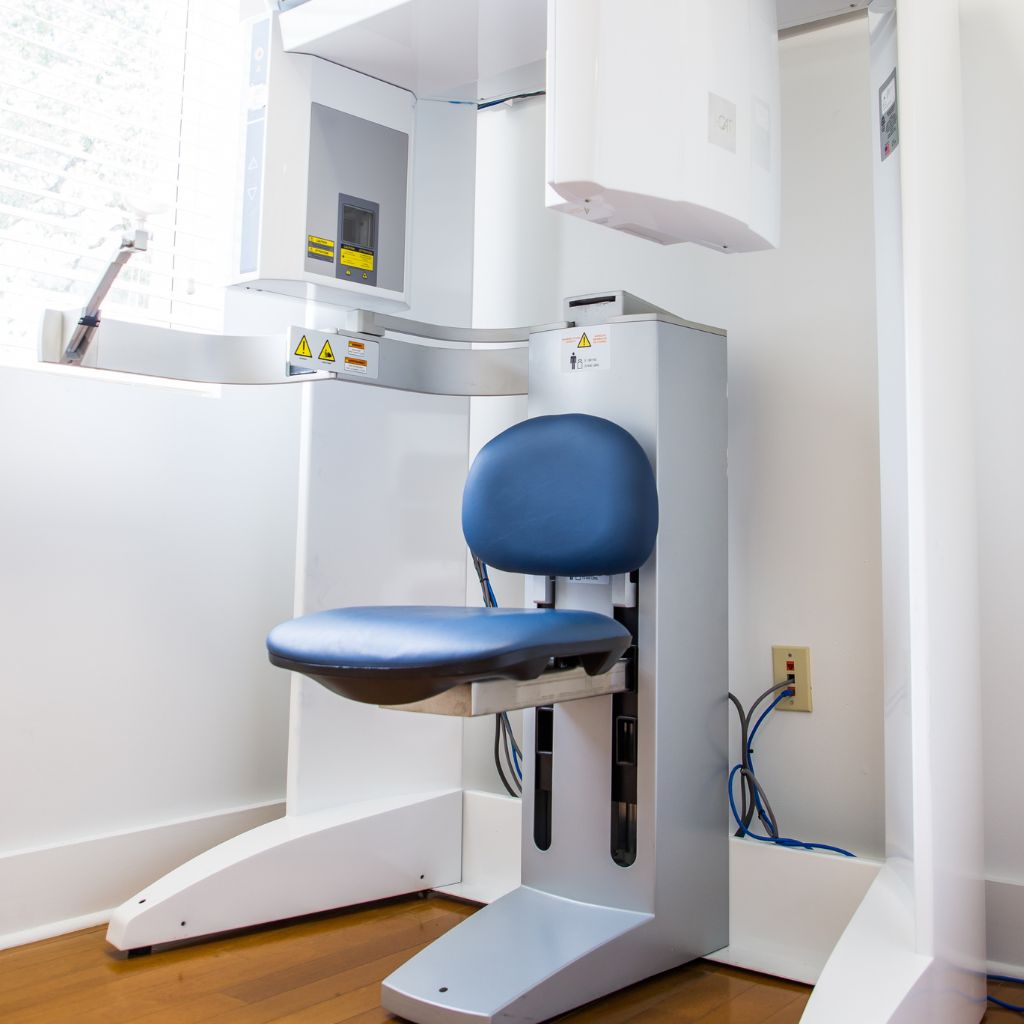
What Is Cone Beam CT Scanning?
Cone Beam Computed Tomography (CBCT) represents the gold standard in dental and maxillofacial imaging. Unlike traditional two-dimensional x-rays that flatten complex oral structures into a single image, CBCT technology creates detailed three-dimensional images of your teeth, soft tissues, nerve pathways, and bone structure with exceptional precision.
The cone beam scanner rotates around your head, capturing hundreds of distinct images from different angles in just seconds. These images are then compiled using sophisticated software to create a comprehensive 3D model of your oral and maxillofacial region. This revolutionary approach provides Dr. Johnson with unprecedented visualization of your anatomy, allowing for detailed examination from every possible angle and perspective.
What truly sets CBCT imaging apart is its ability to reveal hidden conditions and anatomical details that might go undetected with conventional imaging. This enhanced diagnostic capability enables more accurate treatment planning, reduced surgical risks, and ultimately, better outcomes for our patients.
Dr. Douglas L. Johnson, DMD
With over 25 years of specialized experience, Dr. Johnson provides exceptional care using the latest techniques and technology.
Dr. Johnson is a board-certified oral and maxillofacial surgeon who completed his education at the University of Pittsburgh School of Dental Medicine and advanced training through fellowships at prestigious institutions. His extensive training and experience allow him to perform complex procedures with precision and care.
At our state-of-the-art facility, we prioritize your comfort and results. Dr. Johnson and his specially trained staff are committed to providing you with the highest standard of care in a welcoming environment.

Benefits of Advanced 3D Imaging
Advanced 3D imaging provides significant advantages over traditional radiography, starting with dramatically enhanced diagnostic accuracy. The detailed visualization of all oral and maxillofacial structures allows Dr. Johnson to identify conditions that might be missed on conventional x-rays, ensuring more complete diagnosis and appropriate treatment planning. This precision directly translates to improved surgical outcomes by enabling meticulous pre-surgical planning and reducing procedural complications.
For patients, this technology means significantly reduced radiation exposure compared to traditional CT scans—up to 10 times less—while still providing superior diagnostic information. The non-invasive, comfortable scanning process takes just seconds to complete, eliminating the discomfort associated with traditional dental impressions for many procedures.
Perhaps most importantly, 3D imaging enhances communication between doctor and patient. The detailed visualizations allow Dr. Johnson to clearly explain your condition and treatment options, helping you make fully informed decisions about your care. This educational component creates greater confidence and reduces anxiety about upcoming procedures, knowing that your treatment plan is based on the most complete diagnostic information available.
Our Reviews
Why Patients Choose Us
State-of-the-Art Technology
Our practice has invested in the latest generation of cone beam CT technology, providing superior image quality while minimizing radiation exposure. This commitment to technological excellence ensures you receive the most accurate diagnostics available in modern oral surgery, forming the foundation for successful treatment outcomes.
Specialized Expertise
Dr. Johnson combines advanced technology with specialized surgical expertise to interpret imaging results with exceptional clinical insight. This integration of technology and clinical knowledge means your diagnostic images aren’t just viewed—they’re comprehensively analyzed to develop the most effective treatment approach for your specific condition.
Patient-Centered Approach
We believe in making advanced technology accessible through clear explanation. Dr. Johnson takes time to review your imaging results with you, explaining findings in understandable terms and answering questions thoroughly. This educational approach helps you feel more confident and informed about recommended procedures.
Frequently Asked Questions
Is 3D Imaging Safe?
Yes, our cone beam CT scanner uses significantly less radiation than traditional medical CT scans—up to 10 times less. The radiation dose is only slightly higher than conventional dental x-rays while providing vastly more diagnostic information. We follow ALARA principles (As Low As Reasonably Achievable) to minimize exposure while obtaining necessary diagnostic information. Protective equipment is also used when appropriate for additional safety.
Will My Insurance Cover 3D Imaging?
Many insurance plans provide coverage for advanced imaging when medically necessary for diagnosis and treatment planning. Coverage varies by provider and plan specifics. Our experienced office staff will verify your benefits prior to imaging and discuss any potential out-of-pocket costs. We believe in transparent financial communication and will ensure you understand any costs associated with your diagnostic imaging before proceeding.
How Long Do Dental Implants Last?
With proper care and maintenance, dental implants can last a lifetime. The implant posts themselves have a success rate of over 95% at 20+ years. The visible portions (crowns, bridges, or dentures attached to the implants) may need replacement after 10-15 years due to normal wear, but the integrated implants typically remain stable for life, making them a permanent solution to tooth loss.
Visit Us in St. Augustine
St. Augustine Oral & Facial Surgical Center is ideally situated at 1301 Plantation Island Dr. Suite #101, St. Augustine, FL 32080, making us easily accessible for patients throughout St. Augustine and surrounding communities.
- 1301 Plantation Island Dr. Suite #101, St. Augustine, FL.
- (904) 460-0505
-
Mon - Thur: 9 AM to 4 PM
Friday: 9 AM to 2 PM
Saturday & Sunday: Closed